Cervical Spine Geometry Correlated to Cervical Degenerative Disease in a Symptomatic Group
Wiegand R, Kettner NW, Brahee D, Marquina N
Research Associate,
Logan College of Chiropractic,
Chesterfield, MO 63006, USA.
rwiegand@logan.edu
OBJECTIVE: To investigate whether a statistical correlation exists between lateral cervical geometry and cervical pathology, as identified on neutral anteroposterior (AP) and lateral radiographs within a symptomatic group; describe the cervical pathology and determine its location and frequency; and identify the subject’s age, sex, and chief complaint.
SETTING: Department of radiology at a chiropractic college.
METHODS: One hundred eighty-six consecutive pairs of AP and lateral cervical radiographs were reviewed for pathology. A 5-category severity scale was used to describe degenerative joint disease, the most common pathological finding. The subject’s age, sex, and symptoms were recorded. Geometric analysis was focused on vertebral position, alignment, and gravitational loading acquired from the neutral lateral cervical radiograph.
RESULTS: Regression and discriminant analysis identified 5 geometric variables that correctly classified pathology subjects from nonpathology subjects 79% of the time. Those variables were:
(1) forward flexion angle of the lower cervical curve;
(2) gravitational loading on the C5 superior vertebral end plate;
(3) horizontal angle of C2 measured from its inferior vertebral end plate;
(4) disk angle of C3; and
(5) posterior disk height of C5. Degenerative joint disease was the most common pathological finding identified within discrete age, sex, and symptom groups.
CONCLUSION: We identified 5 geometric variables from the lateral cervical spine that were predictive 79% of the time for cervical degenerative joint disease. There were discrete age, sex, and symptom groups, which demonstrated an increased incidence of degenerative joint disease.
From the Full-Text Article:
Discussion
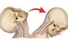
We hypothesized that a correlation may exist between cervical geometry and the presence or absence of cervical degenerative joint disease. Our results identified 5 geometric variables that in a linear combination are predictive 79% of the time of the presence of degenerative joint disease. These variables have the individual and combined effect of anterior head translation. However, the weighted significance of the individual variables within the linear equation describing the likelihood of pathology demonstrated the combined effect was more complicated than simple additive translation.
Anterior head translation, resulting from diminished or buckled cervical configurations, produces abnormal loading, increased mechanical stress, and unbalanced pressure gradients onto the anterior cervical column. [7] These unbalanced pressure gradients, when directed on the skeleton, are known to cause remodeling as described by Wolff’s law. Pressure gradients are also associated with the generation of biopotentials, including piezoelectricity and streaming potentials. [14] These local electromagnetic fields affect the orientation and deposition of bone. Subsequently, metaplasia of fibrocartilaginous tissue into osteophytes and other degenerative findings will occur as physiological responses. [15]
The gender ratio of our 186 subjects was 2.05:1 female subjects to male subjects. The gender ratio within the degenerative joint disease group was 2.13:1. From this data, female subjects had a 4% increased likelihood of degenerative joint disease compared to male subjects. However, our data demonstrated that the age of onset of degenerative joint disease was gender-dependent. Male subjects displayed an 80% increase in the incidence of degenerative joint disease in the age group of 31 to 40 as compared to female subjects of the same age group; female subjects demonstrated an 81% increased incidence of degenerative joint disease in the age group 51 to 60 as compared to male subjects. These results might suggest that in male subjects 31 to 40, adverse mechanical factors are dominating, while in female subjects 51 to 60, hormonal factors are dominating. Only 2 of the 8 subjective complaints, upper extremity paresthesia and shoulder pain, presented with a significantly higher incidence of degenerative joint disease, 60% and 64%, respectively, and these were female-dominant findings.
Our study has important limitations. One is that the same sample was used to estimate both the correct pathology classification percentage and the parameters of the discriminant function. A study needs to be conducted using only the parameters of the discriminate function. This would confirm the reliability of the discriminant function. Another limitation is the small sample size. Clearly, a larger sample needs to be collected across a broader range of cervical pathology, cervical geometry, and age. This would provide a better understanding of the statistical correlation with respect to the frequency of cervical degenerative joint disease, symptoms, and gender. Ultimately, this improved design would enhance our confidence in the stated conclusions. In addition, the utilization of bone densitometry and magnetic resonance imaging in a future study may identify early increases in bone density associated with pathomechanically induced unbalanced pressure gradients.
Conclusion
This study correlated cervical geometry and pathology. We reported (1) in combination, 5 biomechanical variables that correctly classified subjects into pathology and nonpathology groups 79% of the time; (2) the presence of a gender bias in the incidence of degenerative joint disease in specific age ranges, male subjects 31 to 40 and female subjects 51 to 60; (3) an increased incidence of degenerative joint disease associated with upper extremity paresthesia and shoulder pain; and (4) C5-6 was the most frequent location of cervical degenerative joint disease. If the correlation of the 5 geometric variables with joint pathology is supported by further study, the opportunity to predict risk factors for cervical degenerative joint disease is available using cervical radiography [13].


